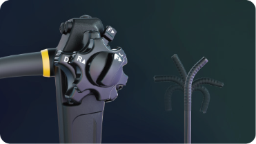
Acquiring high-resolution visual data from within the human body allows clinicians to perform minimally invasive diagnostics and interventions with extreme accuracy. A modern endoscope operates by transmitting light through fiber optics or LED systems to illuminate internal cavities, while a camera sensor captures real-time imagery. COMEN develops these sophisticated imaging systems to ensure that surgeons have a clear, unobstructed view of anatomical structures. By integrating advanced digital processing, every medical endoscope they produce converts light signals into high-definition video, enabling medical teams to identify pathologies that would otherwise remain hidden without major surgical incisions.
Light Transmission and Illumination
Effective visualization begins with the delivery of powerful, cool light to the target area. Manufacturers engineer each endoscope with specialized light cables that prevent thermal damage to delicate internal tissues while maintaining maximum brightness. This illumination is critical for the camera sensor to distinguish between healthy and diseased membranes. Since a medical endoscope must function in a fluid-rich environment, the light source is often calibrated to penetrate various densities, ensuring that the resulting image remains crisp and professional throughout the entire length of a clinical procedure.
Image Capture and Signal Processing
Once the internal site is illuminated, the distal tip of the device captures the visual field using a micro-CCD or CMOS sensor. They design the internal circuitry of the endoscope to transmit these electronic signals through the insertion tube to a video processor without loss of detail. This processor then enhances the contrast and color balance of the footage. A high-quality medical endoscope relies on this rapid data transfer to provide a lag-free experience for the operator, which is essential for coordinating hand-eye movements during complex endoscopic surgeries or biopsies.
Mechanical Articulation and Utility
Control over the direction of the camera is managed through a series of internal wires that allow the tip to deflect in multiple directions. They build each endoscope with ergonomic handles that feature control wheels, allowing the physician to maneuver through tight anatomical curves with ease. Furthermore, a medical endoscope often includes dedicated channels for air, water, or surgical instruments. These integrated pathways enable the clinician to clear the lens of debris or perform minor therapeutic tasks, such as tissue sampling, while maintaining a continuous visual connection to the site.
Reliable internal imaging technology has become a fundamental requirement for modern gastroenterology, urology, and respiratory medicine. Through the combination of fiber optics, digital sensors, and precise mechanical engineering, they provide the healthcare sector with tools that enhance diagnostic speed and patient safety. These technological developments ensure that medical facilities can offer less invasive options for their patients, resulting in faster recovery times and more accurate clinical outcomes across various medical specialties.
